Transmission Electron Microscope (TEM):
This ultra-advanced equipment (LVEM25) offers valuable visualization of the inner structure of a cell sample, including crystal structure and morphology. TEM provides students and faculty members an in-depth understanding of the subject matter related to biological samples and provides skills to students that are needed to be competent in several educational research programs upon graduation. The LVEM25 is a low voltage electron microscope that offers a high-contrast, high-throughput, compact imaging solution with nanometer resolutions that has all the benefits of low voltage microscopy with no limitations. This instrument provides researchers and students with highly advanced structural views, calculations, and structural predictions of samples in teaching and research. The integration of this technology will be very effective to involve students in innovative teaching and research in STEM disciplines.
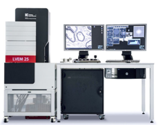
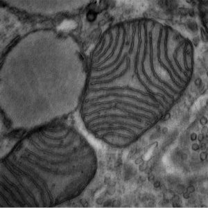
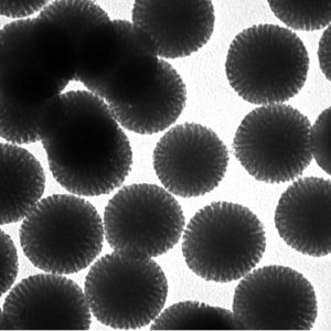
It can also be used to study the activities, functions, properties, and organization of cells and the analysis of fine structures of a biopsy. This specific microscope has the added benefit of being able to work with conventionally prepared thin sectioned materials and doesn't’t require staining of samples. This provides images without the side effects often encountered with staining artifacts.
For equipment use, please contact VITAL STEM Imaging Exploration Center. Also, please review our equipment usage policy prior to use of this equipment.
Micro-Computerized Tomography (CT) Scanner:
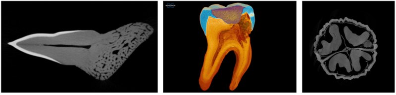
Micro-computed tomography is used to provide X-ray imaging in 3D with significantly increased resolution. The SkyScan1275 is a micro-CT that is specially designed to produce 3D high-speed imaging without compromising image quality. Using new X-ray source technology and efficient flat-panel detectors, the micro-CT reduces scan times down to a few minutes for routine sample scanning at the push of a button. Being able to study the 3-dimensional (3D) structure and morphology of tissues with high-resolution imaging is becoming increasingly important in biomedical research. Micro-computed tomography generates scans similar to CT scans performed in hospitals but on a smaller scale and offers a greatly enhanced resolution.
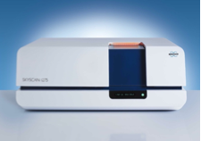
Micro-CTs are often used to evaluate bone structure as it offers excellent cross-sectional images of skeletal structures. For example, it can be used to study bone diseases such as osteoporosis and evaluate the efficacy of therapeutics such as bisphosphates. In addition to bone research, micro-CT is commonly used to study cancer biology. With the development of new contrast agents, micro-CT scanning of soft tissue is an emerging area for this modality, particularly in the area of cardiovascular and pulmonary research.
For equipment use, please contact VITAL STEM Imaging Exploration Center. Also, please review our equipment usage policy prior to use of this equipment.Virtual Reality Pods and VITAL Research Lab
Using advanced VR technologies, students can view the world of Chemistry, Biology, and Forensic Science through a lens that they could not see before. For example, students are able to visualize how the 3D structure of chemical compounds and a protein is represented.
Virtual Reality Pods and VITAL Research Lab
Using our VR facility, students and faculty members can themselves be part of a 3D environment and can get very close to molecules, proteins, the cellular world, and so much more. This advanced VR technology will help our students to be more confident, more engaging, and more involved in many of their classroom topics; and as a result, the rate of their success in many STEM courses will increase.
Currently, more than 7 courses from Chemistry, Biology, Forensic Science, and Environmental Science have already adopted VR classroom teaching and we are hoping more and more courses will adopt these VR teaching technologies.
For equipment/ VR Pods use, please contact VITAL STEM Imaging Exploration Center. Also, please review our equipment usage policy prior to use of this equipment.
Visitors
With VR Halo Classroom, VITAL Research lab, and multiple VR Pods/ stations, our STEM Imaging Exploration Center is located in Hubert-D Building, 3rd Floor (Lab #442, # 444).
To schedule a visit using the contact information below:
|
Anthony Palacios, B.A.
VR Immersive Technology Research Technician
Email: palaciosa@savannahstate.edu
Tel: 912.358.3825
|
Manoj Prasad, Ph.D.
Director, VITAL Title III Program
Email: Prasadm@savannahstate.edu
Tel: (912) 358-4448
|